Oral disease
may present as
- Mucosal surface lesions – white, red, brown, blistered, verruciform
- Swelling at an oral subsite – lip/buccal mucosa, tongue, floor of mouth, palate and jaws
- Symptoms related to teeth – pain, mobility
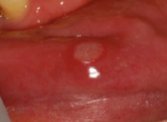
Ulcerated lesions

- Usually traumatic or immunological (aphthous)
- Less common – bacterial, fungal, immune related e.g. IBD
- Residual appearance of a blistering condition
- Need to exclude oral malignancy
- Consider persistence vs episodic recurrence
- Mucosa turns over < 10 days
- If >2 weeks = biopsy
Traumatic ulcers
- Mechanical, biting, thermal, radiation, chemical
- Inflammation, pain, redness, swelling
- Natural ulcer often yellow-white fibrinous exudate
- Address cause, ensure healed in 2 weeks
- If the ulcer persists beyond this period, referral to an oral and maxillofacial surgeon should be undertaken.
Aphthous ulcers
- Recurrent Aphthous Stomatitis common
- affects 20–50% of the population
- aetiology of RAS is unknown
- Occurs in non-keratinised oral mucosa (buccal mucosa, floor of mouth, vestibule of the lips, soft palate and tongue)
- Often missed:
- Crohn’s, Coeliac
- aspirin burn (if sucking on tabs)
- HSV
- EBV
- non-healing
- ?cancer, leukaemia, HIV, syphillus, TB, agranulocytosis
- 3 types
- Minor aphthous ulcers
- oval shaped and <10 mm in size
- frequently last 5–10 days
- heal without scarring.
- Major aphthous ulcers
- variably shaped and >10 mm in size
- can last up to six weeks
- can heal with scarring
- can resemble an ulcer of an early malignancy.
- Herpetiform aphthous ulcers
- numerous 1–2 mm diameter ulcerations
- can occur in clusters of more than 20 at a time which can merge to give larger ulcers.
- They tend to occur in the front of the mouth particularly under the tongue and on the edges of the tongue
- not restricted to non-keratinised oral mucosa
- heal in 1–2 weeks.
- Minor aphthous ulcers

- can be associated with haematinic deficiency or gastrointestinal or rheumatological disease.
- Vitamin B12, folic acid or iron deficiency can also be associated with RAS in a small proportion of patients, and these nutritional markers should be checked and corrected in this subgroup.
- Treatment:
- non pahrm
- Medicated toothpaste without sodium laureth sulphate.
- Antibacterial mouthwashes to reduce secondary infection.
- Avoidance of foods that trigger or exacerbate the ulcers.
- Treatment of an associated condition or underlying cause, for example –
- Any iron or vitamin deficiency should be corrected
- Reduction in stress.
- pharm
- Superficial tissue cauterisation using silver nitrate stick.
- Topical corticosteroids
- hydrocortisone 1% cream or ointment 2–3 times daily after meals
- Hydrocortisone hemisuccinate pellets (Corlan), 2.5 mg 4 times daily
- Triamcinolone (Kenalog): administered 4 times daily
- Betamethasone 0.5-mg tablet dissolved in 15 mL of water to make a mouth rinse, used 4 times daily for 4 minutes each time
- Local anaesthetics benzocaine and lignocaine (lidocaine) to reduce pain
- to stop recurrence
- brush atraumatically (eg, with a small-headed, soft toothbrush)
- avoid eating particularly hard or sharp foods (eg, toast, potato crisps)
- avoid other trauma to the oral mucosa.
- non pahrm

Oral SCC

- Most common oral cancer
- Risk factors: Smoking, alcohol, betel quid (paan)
- oral SCC is found across virtually all age groups (including paediatric, although rarely), and up to 10% of SCCs are diagnosed in patients who do not smoke or drink alcohol.
- most commonly presents as
- Non healing ulcer
- indurated/firm
- irregular margins
- raised/rolled edges
- May have symptoms due to invasion of local structures 🡪 Refer Max Fax
Ulcer on lip
- similar to skin cancer
- presents as a non-healing ulcer on the vermillion of the lower lip
White, red and mixed lesions

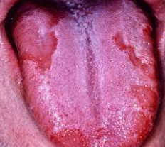
Geographic tongue
- Aka Erythema migrans
- effects approximately 1–3% of the adult population worldwide.
- less common in children.
- may have a family history of geographic tongue or fissured tongue.
- The cause is unknown
- often in patients who have psoriasis. food allergy, allergic contact dermatitis, asthma, atopic dermatitis, reactive arthritis, anaemia, hormonal disturbance, emotional stress and in patients with early-stage of type 1 diabetes.
- differential diagnosis
- Oral candidiasis
- Oral psoriasis (rare)
- Oral lichen planus
- Trauma
- Herpes simplex
- Systemic lupus erythematosus
- Oral leukoplakia
- Appearance changes over several days
- Asymptomatic, reassure, In most cases, it resolves over time without treatment
Black Tongue

- -overgrowth of papillae caused by poor oral hygiene, debility
- -also abx, steroids, tranquilisers
- -brush/scrape black area
- -suck on pineapple piece for 40 seconds then chew slowly, rpt 8 times bd
Median Rhomboid Glossitis

- -central papillary atrophy,loss of lingual papillae
- -usually fungal infection, worse with smoking, dentures, HIV, steroids
- -treat with antifungal lozenges
Frictional keratoses

- Chronic low grade trauma e..g at bite line
- Might appear lighter colour and take on shape/outline
Oral submucous fibrosis

- Fibroelastic change in the oral mucosa
- Potentially malignant
- Stiffness of mucosa, trismus
- Risk factors: South east Asia, betel quid
- characterized by progressive
- restriction of mouth opening
- blanching of the mucosa
- ‘guitar string’ sensation when palpating the buccal mucosa
- depapillation of the tongue
- loss of pigmentation of the mucosa
- Cease contributing factors
- Needs ongoing surveillance
Leukoplakia (diagnosis of exclusion)
- descriptive term for white plaque rather than a microscopic tissue diagnosis
- tissue diagnosis may range from benign (eg hyperkeratosis) to pre-malignant (dysplasia) to malignant (SCC)
- Needs biopsy
Erythroplakia (diagnosis of exclusion)

- refers to a red patch of the oral mucosa or a red lesion that cannot be characterised clinically or pathologically as any other definable lesion or disease
- Red counterpart of leukoplakia
- More likely to be SCC
- Key points
- The vast majority of oral mucosal and jaw conditions are benign and amenable to surgical, medical or dental treatment.
- The possibility of an oral cavity malignancy should always be considered, particularly with the presentation of a non-healing ulcer, a bleeding lesion or an area of mucosa that persistently appears red or white.
- Any persistent ulcer that has been present for ≥2 weeks should be referred to an oral and maxillofacial surgeon or oral medicine specialist for biopsy.
- Leukoplakia and erythroplakia are clinical terms used to describe oral white and red patches respectively that cannot be scraped off and cannot be ascribed to any disease or condition. Referral for biopsy is mandatory as a significant proportion of these lesions will be dysplastic or malignant.
- Any tissue excised from the oral cavity should be sent for histopathological examination as the clinical appearance alone is insufficient to ensure a correct diagnosis
oral cavity
Submucosal swelling
- Mucocele


- Smooth fluid filled lump
- Commonly lips, buccal mucosa
- Often in areas with salivary glands after trauma allows mucus to escape
- Should spontaneously resolve 2-3 weeks
- can occasionally be clinically difficult to distinguish from a minor salivary gland tumour.
- Any mucocele should be excised and sent for pathological examination
- A ranula
- is a mucocele of the sublingual gland
- Treatment involves removal of the sublingual gland (marsupialisation or incision and drainage alone leads to unacceptably high recurrence rates)

- Fibroepithelial polyp
- Outgrowth from mucosa
- Usually exuberant healing following trauma
- Surgical excision
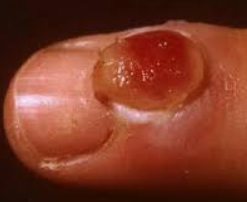
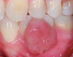
- Pyogenic granuloma
- Red polypoid lesion
- caused by an exaggerated connective tissue response to minor trauma
- bleeds easily
- commonly found in – Gingiva, lateral aspect of tongue, lower lip, buccal mucosa
- Treatment includes surgical excision and removal of the traumatic irritant (eg subgingival plaque)
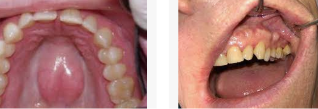
Palate
- Palatal torus, asbcess or cyst, minor salivary gland tumour
- Should all be referred to Max fax
exostoses and tori
- Hard bony protuberances, covered by normal appearing mucosa
- Often asymptomatic
- May see on OPG, consider CT scan
- Doesn’t need particular management unless symptomatic (torus/exostosis interferes with the placement of a removable dental prosthesis (denture) or is growing, or the overlying mucosa is recurrently ulcerated)
Cysts of the jaws
- Usually detected incidentally on OPG
- Periapical cysts most common -inflammatory, develop at the apex of a non-vital tooth